 Dom & Cleo True Colloidal Silver Spray 2oz | Kohepets
Warning: The NCBI website requires JavaScript to operate. Ultrastructural Analysis of Albicans Candida when exposed to silver nanoparticles Roberto Vazquez-Muñoz 1 Department of Microbiology, Center for Scientific Research and Higher Education of Ensenada (CICESE), Ensenada, B.C., Mexico, Miguel Avalos-Borja 2 Advanced Materials Division, Potosino Institute for Scientific and Technological Research (IPICYT), San Luis Potosí, S.L.P., Mexico, 3 Centro de Nanociencias y Nanotecnología, Universidad Nacional Autónoma de México (UNAM), Ensenada, B.C., México, Ernestina Castro-Longoria 1 Department of Microbiology, Center for Scientific Research and Higher Education of Ensenada (CICESE), Ensenada, B.C., Mexico, Designed and designed the experiments: RVM ECL MAB. He performed the experiments: MVR. He analyzed the data: RVM ECL MAB. Reactive/material/analysis tools: MAB ECL. Contributed by Writing of the manuscript: ECL RVM MAB. Associate Data The authors confirm that all underlying data of the findings are fully available without restriction. All relevant data are within the document. Summary Candida albicans is the most common fungal pathogen in humans, and recently some studies have reported the antifungal activity of silver nanoparticles (AgNPs) against some Candida species. However, ultrastructural analysis of the interaction of GNPs with these microorganisms has not been reported. In this work we evaluate the effect of the AgNPs on the C albicans, and the minimum inhibitory concentration (MIC) was found with a fungicidal effect. The IC50 was also determined, and the use of fluconazole (FLC), a fungitic drug, a reduction of cell proliferation. To understand how the AgNP interact with the living cells, the ultrastructure distribution of the AgNPs was determined in this fungus. The analysis of electron transmission microscopy (CT) revealed a high accumulation of AgNPs outside the cells, but also smaller nanoparticles (NPs) located along the cytoplasm. The analysis of energy dispersed spectroscopy (EDS) confirmed the presence of intracellular silver. Of our results it is assumed that the AgNPs used in this study do not penetrate the cell, but release ions of silver that infiltrate the cell that lead to the formation of PN through the reduction by organic compounds present in the cell wall and cytoplasm. Introduction Pulmonary infections are among the main causes of infectious diseases, , with Candida being the most representative model of pathogenic yeasts in humans . Candida albicans is a dimorphic fungus that is present as an important component of normal flora in healthy people, but in conditions of a weakened immune system Candida becomes an opportunistic pathogen after a transition from a pathogen to a pathogen. Candida species are able to form biofilms and are the main cause of mortality in immunocompromises patients, as they cause invasive candidiasis, which is difficult to eradicate due to high resistance to antifungal treatments. In the fight against fungal infections, some antifungal substances have three main disadvantages: limited range of action, self-medication, which can negatively interact with different types of antifungal agents, and resistance of microorganisms, . In addition, despite the improvement of antifungal therapies over the past 30 years, antifungal resistance is still of great concern in clinical practice, and in general new costs of antifungal development Silver has long been recognized as an effective antimicrobial agent even in low concentrations, and recently silver nanoparticles have gained recognition as an antibacterial agent/promising antifungal. Silver is used in clinics to treat pathogenic infections in skin wounds, burns and transplant surgery; however, the chronic use of silver to treat diseases, or take sterile coloids. Silver is eliminated from the human body by the liver and kidneys and apparently not toxic to low concentrations, but at higher concentrations it is considered potentially toxic to human cells, inhibiting protein and DNA synthesis. Because of the importance of silver in health care, recent studies suggest that the AgNPs can be safer to use than ion or colloid silver. Recently, Huh and Kwon defined the term "nanoantibiotics" as nanomaterials with antimicrobial activity and/or for those who improve the effectiveness and safety of antibiotics, of which the ANPs are currently among the most studied –. Possible positive effects on the use of AgNPs are the prevention of the formation of biofilm, anti-inflammatory activity –, antiviral capacity and use in post-burning treatments. AgNP has also been reported to have synergistic interactions with different antimicrobial drugs, which is particularly important because in some treatments the amount of drugs could be reduced along with the concentration of AgNP. However, more research is needed to clarify the safety of the use of GNPs in the treatment of human diseases. In this regard, most of the reports currently point to their use against pathogenic bacteria, but studies on the effect of GNPs against other microorganisms such as fungi, protozoa and viruses are limited. In addition, most studies related to the biological systems of AgNPs focus on the biocid effect of nanomaterials on microbes, and only a limited number of studies explore the interactions and possible mechanisms of action of the AgNPs in microbial inhibition. AgNPs' antifungal activity has been tested against different species of Candida; C. albicans, C. glabrata and C. tropicalis were completely inhibited using AgNPs. In addition, it was reported that the AgNPs damaged the cell membrane structure in C. albicans producing "holes" on the surface of the cells and thus inhibited the lace process. Production of reactive oxygen species (ROS), DNA fragmentation and apoptosis has also been reported. Although the antifungal effect of GNPs is generally known, their interaction with biological systems is not fully documented. Therefore, the objective of this work is to evaluate the effect of a commercial product AgNPs against C. albicans and to determine how the AgNP interact with Candida cells at ultrastructural level; this information will complement existing information and could contribute to generating new antifungal treatments of a wide spectrum. Materials and Methods Conditions of training, means and growth Candida albicans ATCC SC5314 cultivated strain at 37°C in liquid and solid media: YPD (1% yeast extract, 2% peptona, 2% dextrose) and YPD agar plates (2% added bacteriological agar). The cultural media were prepared using distilled water and sterilized by conventional methods. The AgNPs used in this work were Vector Vita Ltd, Novosibirsk. The product is defined as an "Argovit" medication that is grouped silver (AgNPs) functionalized with polyvinylpirrolidone (PVP) and the average reported size of initial cluster particles is approximately 1.5–2 nm. The silver concentration of the product is 12,000 μg/mL.Minimum inhibitory concentration (MIC)The minimum inhibitory concentration (MIC) of AgNPs was determined by a macrodilution test as follows: The cells were cultivated in YPD agar plates for 24 hours, at 37°C. Then, 2–10 colonies were inoculated in the YPD broth and the cell density was adjusted using a spectrophotometer. Immediately, cultures were exposed to different treatments (AgNP, FLC and AgNPs -FLC) and incubated at 37° C for 24 hours at 250 rpm (Orbit Environ Shaker). The cultures of three cell density were exposed to different silver concentrations, and after treatment, the cells were inoculated in YPD agar plates and allowed to incubate for 24 hours to 37°C. To determine the combined effect of AgNPs with a commercial antifungal product, using the method described above, the medium-maximal inhibitory concentration (IC50) was determined for AgNPs and fluconazole (FLC). The standard absorption growth curve (abs) vs. cell density in YPD was performed in λ = 520 nm in a Jenway spectrophotometer 6505 UV-Vis; all measurements were performed in triplicate, and the standard deviation was calculated (σ). Sample preparation for transmission electron microscopy (TEM) For microscopic analysis, cultures exposed to the determined MIC of AgNPs were first observed after 24 hours of incubation under bright field microscopy, using a Zeiss Axiovert 200 M. After that, cultures were prepared for the analysis of transmission electron microscopy (TEM). During the preparation process, the samples were centrifuged at each step of the protocol, 5 min to 1500 rpm for the fixing and dehydration processes and 10 min to 3000 rpm during the infiltration. Pellets were fixed with 2% glutaraldehyde in 0.05 M of sodium phosphate for 30 minutes at room temperature. After the fixing cells were washed with sodium phosphate and fixed post with 1% OsO4, for 2 hours to 4°C. Subsequently, the samples were dehydrated in ethanol series (15, 30, 50, 75 and 100%) for 2 hours at each step. Then the samples were infiltrated into the resin/ethanol series (20/80, 40/60, 60/40, 80/20) for 3 hours at each step and stayed overnight to the 100% Spurr resin. Finally, the pellets were placed in the clips previously coated with Teflon spray and "sandwiched" in the 100% Spurr resin for polymerization at 60° C for 24 hours. After the cooling, the lids were separated and examined under a stereo microscope to cut resin pieces containing pellets. Polymerized pellets were mounted in resin blocks and selected in an ultramicrotome Leica Ultracut R. The 70 nm sections were mounted in mesh copper grids in form/carbon 75 and analyzed under TEM (Hitachi H7500 operated at 100 keV and spot size 5). The sections were examined without postscripts for better detection of silver. High-resolution transmission microscopy (HRTEM) To determine the chemical characterization of the ultratin C. albicans sections and crystal-inographic analysis of silver, the samples previously scanned under TEM were examined under microscopy of high-resolution transmission electrons (HRTEM) (Tecnai F30 operated at 300 keV and spot 6 size). The analysis included Scanner Transmission Microscopy (STEM) and Dispersive Energy X-ray Spectroscopy (EDS). Results It is known that the AgNPs are highly volatile, and previous studies have shown that the size of the AgNP could influence their antimicrobial activity. Therefore, NPs used to treat albicans C. were examined under TEM to determine the range of form and size at the time of the experimental procedure. It was found that the AgNP were mainly quasi-spheric, although other forms were also detected (). The size range was 3–60 nm () before and after the application, being different from that reported by the authors. The elemental character of nanoparticles was also determined and the analysis EDS confirmed the presence of silver (). The presence of copper in the obtained results could be due to the grid we used; however, the silicon signal () could be a trace component of the commercial product we used. A) AgNPs electronic transmission micrograph functionalized with Polyvinylpyrrolidone (Vector-Vita Ltd., Russia), B) nanoparticle histogram measurements, C) ANPs's dark field annular images (HAADF), D) Typical Particle EDS analysis along the stroke indicated by a white line in (C). The minimum inhibitory concentration (MIC) of the AgNPs depends to a large extent on the initial inoculum and the size/form of the NPs; therefore, the MIC was determined for cultures of different cell density (). In all cases, MIC was equivalent to the minimum concentration of fungi (MFC). After 24 h of treatment exposure, the samples were re-induced in YPD agar plates, and after 24 h of incubation no growth was detected in cultures exposed to AgNPs. In addition, since the combined use of an antimicrobial drug with AgNPs reduces the amount of the drug used to inhibit microorganisms, the efficacy of the combined effect of AgNPs with a fungitic drug was determined; we use fluconazole IC50 (FLC) and AgNPs IC50 against different concentrations of Candida cells. Using 2.5×10⁶ cells/mL, the AgNPs IC50 was 60 μg/mL and 125 μg/mL for FLC; for 1×10⁴ cells/mL the IC50 was 18 and 31 μg/mL for AgNPs and FLC, respectively (). After 24 h of incubation using the combined IC50 of AgNPs -FLC against Candida (1×10⁴ cells/mL), the optical density of culture was lower than that of control and culture only exposed to FLC IC50 (). The optical density by a higher concentration of FLC (10× IC50 = 300 μg/mL) with the same AgNPs IC50 was similar to the use of 300 μg/mL of FLC alone (). When the AgNPs MIC (42 μg/mL) was used in combination with the FLC IC50 (31 μg/mL) and with 300 μg/mL of FLC, Candida was completely inhibited (). The optical densities recorded using these concentrations were due to the absorption of NPPs. To determine cell viability, samples were inoculated in YPD agar plates and growth was not recorded using 42 μg/mL silver concentration (). Liquid cultures were exposed to FLC's IC50 and various combinations of AgNPs-FLC.A) A representative image of a culture to determine the IC50 subcultures of AgNPs and FLC; B) of the combined effect of the IC50 of AgNPs (18 μg/mL) and two concentrations of FLC subcultures; C) of the combined effect of the AgNPs MIC (42 μg/mL) and two concentrations of FLC. In B-C, FLC IC50 (31 μg/mL) was used in the inoculation on the left side of the plate and FLC 300 μg/mL on the right side. Table 1Inoculum initial and optical density (D.O) of culture (λ = 520)AgNPs (μg/mL silver)4.3×10⁷ cells/mL, O.D = 0.7066002.5×106 cells/mL, O.D. = 0.0821501×10⁴ cells/mL, O.D. = 0.04042 Table 2 C. albicans TreatmentConcentration (μg/mL)2.5×106 cells/mLAgNPs150, 120, 90, 60, 42, 18FLC300, 125, 63, 31,151×104 cells/mLAgNPs90, 60, 42, 18, 6FLC125, 63, 31, 15, 7The cultures of control and cultures exposed to equivalent concentrations of the polyvinylpirrolid stabilizer agent It was determined that PVP does not have inhibitive effects on Candida cells, as there was no difference between the growth of cultures treated with PVP (abs = 2.207, σ = 0.042) compared to control cultures (abs = 2.203, σ = 0.055). The growth in the media of YPD agar plates was also similar in both cases (not shown images). Using bright field microscopy, cultures were first examined to determine cell morphology. Normal cell growth was observed in the control cultures () while in people treated with AgNP cells were observed agglomerated, with nanoparticles around C. albicans (). TEM analysis revealed that cells in control cultures and cells exposed to GNPs did not show serious damage, such as well formation or cell wall rupture (). On the other hand, AgNP was found in most cases surrounding the cells examined (). The chemical analysis was carried out throughout an area, including the cell wall and part of the inside of the cells (). Samples were examined to determine the elemental nature of the particles surrounding the cells. No silver was found in the control samples. Noise was only detected (), while in the cells exposed to GNPs, the presence of silver was clearly confirmed (). A closer inspection in the cytoplasmic area revealed small points of approximately 10 nm in size (); therefore, a chemical analysis was performed to corroborate the nature of these NPPs. The EDS analysis confirmed the presence of silver (), and the crystalline analysis using HRTEM confirmed the occurrence of intracellular silver crystals (). TEM analysis clearly showed that near the extracellular accumulation of AgNPs, a high accumulation of very small particles was present on the cell wall and part of the cytoplasm (). The size of these particles was in the range of 6-12 nm in diameter (), although other sizes were also present in cytoplasm. It is interesting to note that on the border between cytoplasm and cell wall is not clearly defined as in , so it may be possible that the AgNPs interrupt these structures and can cause serious damage in higher exposure times.A, B) Cells of control cultures observed under bright field optical microscopy and TEM respectively; C, D) The cells exposed to silver nanoparticles were a clustered field and surrounded by bright microscopying as optical observatotics. Black arrows indicate that the Candida cells and white arrows indicate AgNPs aggregation. A, D) High-angle dark field images (HAADF) of analyzed cells; (B, E) EDS analysis that show the absence of silver in (A) and the presence of silver in (D); (C, F) linear EDS spectrum (A) and (D), respectively. The red line in A and D indicates the transect where the chemical analysis was performed. A) Image HAADF in which the internal ANP analysis was performed, B) Closer view of the external internal particles, C) Amplified image of the internal particle analyzed (D-F) Images of internal particles analyzed, G) Analysis of EDS that shows the presence of silver, H) Variation of Ag and Os along a line of trace in neighboring points near the Eparticle indicated by a yellow arrow in the outer figs The closed area in (F) indicates the area where the chemical analysis was performed, and the image in the upper corner is the pattern of diffraction of analyzed particles, confirming the presence of crystalline silver. Nano silver particles shows (111) planes with space of 0.24 nm. A, B) Sections of cells in which extracellular agglomeration of GNPs coincided with the accumulation of smaller GNPs on the cell wall and cytoplasm, C) Distribution of size of intracellular GNPs. Debate According to the definition of fungitic versus fungicide effect, in our AgNPs study they were found to present a fungicidal effect, as cultures treated with MIC could not recover after treatment, even after inoculation in the middle of fresh culture in successive subcultures. AgNPs antifungal properties against C. albicans have been shown in some other studies, although MIC's reported values are different from those we find in this work, , , , , , , , . Such differences can be due to the nature of the particles used, the size difference being especially important. It is known that the size and shape of metal nanoparticles influence their chemical, optical and thermal properties. Therefore, the size and other important features could also modify the antimicrobial properties of nanoparticles. For example, the bactericidal properties of GNPs differently against E. coli were found dependent on form, and the size-dependent toxicity was reported in alveolar macrophages. The stabilizing agent can also influence the antimicrobial activity of the NPs, as less MIC of the AgNP stabilized against Candida spp was used, comparing the antifungal activity with the unstabilised AgNPs. In our study, the AgNPs are functionalized with PVP, which is considered biocompatible and did not exercise inhibition in Candida. It is important to mention that in our study, it was observed that the AgNPs (at concentration above 5 μg/mL) slowly sedimented into the YPD broth. The sedimentation was observed in the form of precipitation of particles at the bottom of the test tubes, while no sedimentation was observed in distilled water. Sedimentation could affect the effectiveness of GNPs; therefore, this is another factor that should be evaluated by exposing microorganisms in different conditions of the media of liquid culture. We explore the synergistic effect of AgNPs−FLC against Candida and find that the combination of both agents significantly reduced cell viability, similar to the findings of an earlier study. The use of AgNPs in combination with antimicrobial drugs can be beneficial in the clinic, as the amount of both agents can be substantially reduced and thus avoid adverse effects caused by some drugs and the possible toxic effects to chronic exposure to silver. Some studies have reported that AgNPs exhibit synergistic effects with antibacterial drugs such as amoxicillin, ampicillin, kanamicin, erythromicin and cloranphenicol, penicillin G, clindamycin and vancomycin. However, antagonistic interactions with amoxicillin and oxacillin were detected in a Staphylococcus aureus strain resistant to methicilline and with chloramphenicol in Pseudomonas aeruginosa . Studies on the combined effect of AgNPs with fungicides can be particularly useful as we use FLC, which is considered among fungitic agents. In addition, it is known that even fungic agents have limited action in immunosuppressed patients; therefore, a synergistic effect of antimicrotic drugs can be particularly useful in some clinical cases. The antimicrobial mechanisms of nanomaterials are not fully understood, but it is proposed that when they come into contact with cells, they cause the production of reactive oxygen species (ROS), cell membrane disorder, mitochondrial damage and DNA damage, among others. Ultrastructural studies on microbial interaction with nanostructured material are scarce; this information could provide a better understanding of the NPP mode. There are several ultrastructure-level reports on the interaction of the AgNPs with bacteria; however, some of them present low magnification images that make it difficult for NPs to localize. However, the effectiveness of the AgNPs against bacteria is clearly demonstrated, and in fact, the AgNPs were effective against E. coli, with cells that showed the formation of "pits" on the cell wall. The silver nanoparticles were found to accumulate in the bacterial membrane, and some of them were reported to successfully penetrate the cells. Similar results were found in E. coli and V. cholera; it was established that the AgNP caused changes mainly in the morphology of the cell membrane, producing a significant increase in its permeability, thus affecting proper transport through the plasma membrane, leading to cell death. They also reported that the silver NPs with small diameters penetrated the cells. As mentioned above, the effect of the AgNPs on fungal species is scarce, but the MIC to inhibit Candida spp has been reported by several authors. However, it is necessary to clarify how NPPs interact with fungal cells to generate information that could be useful in developing new clinical techniques for NPP applications. It was recently reported that the effects and possible mechanisms of GNPs in C. albicans were the production of ROS and the nuclear fragmentation leading to cell death. However, as far as we know, there are very few studies that report at an ultrastructure level the effect of the AgNPs with fungal cells. It was said that the C. Albicans exposure to AgNPs produced significant changes in the membrane, the formation of "pits" on the cellular surface, and finally the formation of pores and cell death. In fact, they presented TEM micrographs, but locating NPs outside or inside the cell is not possible due to low magnification images. In our study, Ag NPs were found around C. albicans cells, similar to the results found in bacteria, ; also, AgNPs were found not specifically distributed within the cell cytoplasm and in different regions of the cell wall, but no serious damage was observed in the structure, which differs from a study that reports the presence of "holes" in the Candida cell wall when it is treated with Agdidas. Another study reports the interruption of the cellular wall and the cytoplasmic membrane in the neoforms of Cryptococcus treated with AgNPs in a 72-hour exposure time. From our analysis TEM was clear that the accumulation of small AgNPs on the cell wall coincided with the accumulation of extracellular NPs, which strongly suggests a dynamic release of silver ions (Ag+) by the adjacent AgNPs that actively penetrate the cell and lead to the intracellular biosynthesis of AgNPs. The results of the HRTEM analysis of intracellular NPs show a set of lines spaced by 0.24 nm, which is consistent with (111) silver planes (). The gradual release of Ag+ by AgNPs has been suggested earlier so that the internalization of Ag+ in the cell can be of particular relevance, as they can act as an embryo increasing the duration of antimicrobial effects. This is of paramount importance, as the AgNPs are shown to inhibit the formation of biofilm, thus avoiding stronger and larger infections, or could represent a greater durability of the medical devices with AgNP contents. However, despite the clear antimicrobial properties of the AgNPs, their potential use in the clinic should be carefully evaluated as there is a lack of basic knowledge about the potentially different antimicrobial properties of the AgNPs that can vary depending on many factors, including the synthesis method, size, shape, functionalizing agent, application method etc., and also their interaction in more complex systems such as plants, animals and humans. ConclusionsThe results obtained in this study complement the existing research on the potential use of nanomaterials in biomedicine. The exgicidal capacity of the AgNPs functionalized with PVP was determined. It was clearly shown that although no dramatic damage was observed to the fungal cells, at least after the 24h exposure time, no cell viability was recorded. Another important result was to discover that the action mode of the AgNPs is to add outside the fungal cells, freeing ions of silver and thus inducing cell death through the reduction process resulting from the interaction of cellular components with ion silver. Thank you. Thank you. Rosa Mouriño (CICESE) for providing Candida strain, Dr. Nina Bogdanchikova (CNyN-UNAM) for providing nanoparticles of silver, Naidy Pinoncely, Isadora Clark (CICESE) and Jennifer Eckerly (IPICYT) for technical assistance and Dr. Nicolas Cayetano (LINAN-IPICyT), for help with HRTEM work. Statement of funding This work was partially supported by a SEP-CONACyT grant (CB2011/169154). The authors also thank CONACyT for a scholarship to Roberto Vazquez-Muñoz. Funders had no role in the design of study, data collection and analysis, the decision to publish or the preparation of the manuscript. Availability Authors confirm that all the underlying data of the findings are fully available without restriction. All relevant data are within the document. ReferencesFormats: Share , 8600 Rockville Pike, Bethesda MD, 20894 USA
All the things that enrich. Why? How to make a colloidal silver bag (+ How I healed cervicitis Naturally when antibiotics didn't work!) Learn about healing powers of colloidal silver, along with why and how to make a colloidal silver bag for women's problems such as yeast infections, bacterial vaginosis, and cervicitis. Also, how I cured my cervicitis naturally after 2 antibiotics didn't work! Disclaimer: This post is intended for information purposes only and is not a substitute for the advice and care of a qualified medical professional of your choice. I've done enough research that I feel I should have letters behind my name, but unfortunately, I don't. So please do your own research and consult your doctor(s) when making health decisions. In mid-January 2019, I was in the middle of a health crisis involving as much as . For a few days, I had felt a heavy and dragging feeling in my pelvis, almost as if something was going to come out of my vagina. As I progressed, I noticed that I lost the use of my pelvic soil muscles and couldn't make a Kegel. Finally, I had to examine myself. So, I sat in the toilet and gently put my index finger on my vagina. Before I had entered my first knuckle (less than an inch), I could feel my cervix. Thinking that gravity/feeling might have something to do with it, I called my husband nervously and put myself on the bathroom floor so I could check myself out too. I felt what I felt, and we decided that I needed to get medical attention right away. Diagnosis of my cervicitis In the ER, the doctor performed a pelvic exam and immediately commented: "You have a rabies cervicitis." Never having heard the term "cervicitis", we, of course, asked what it meant. "Basic inflammation or infection of your cervix," the doctor replied. Have you noticed any white downloads? Any pain with sex? Does it bite you? " I answered NO to all your questions. The only symptom was this heavy and dragging feeling on my pelvic floor and the inability to properly use my pelvic soil muscles. Everything that was down there felt so bad. It was really scary. We learned that the causes of cervicits are: bacterial vaginosis, gonorrhea and chlamydia sexually transmitted diseases, and tricomoniasis. Of course, the doctor tested all these infections and I was negative for ALL. Until today, the cause of my cervicitis remains a mystery. He had none of the typical symptoms and any of the risk factors. The only thing I can guess is that I was low, along with PCS and pelvic soil dysfunction, that my space in the belly had become susceptible to bacteria. That I had no symptoms, except for the feeling that my cervix was going to fall, it's also a mystery. However, I was given an injection of antibiotics and a prescription for antibiotic pills, so 2 different and strong antibiotics, to resolve cervicitis. After a week, my cervix was still swollen and tense. (We've been checking it daily.) So, I consulted my functional doctor, recommended a silver colloid bag. Besides, I consulted my midwife. Surprisingly, I had never heard of playing with colloid silver, but he said it looked like a sound advice. He recommended inserting specific vaginal probiotics every day, in addition to the imbeciles. Why the Colloidal Silver should be in every Medical Cabinet So what is it?Colonial silver is a natural antimicrobial consisting of silver particles suspended in water. It has been used literally for ages, traced back to medieval times, ancient Greeks and Romans! If the colloidal silver is not part of your medicine cabinet, it must be. And here's why... it has powerful antimicrobial properties. You can even kill antibiotic-resistant superbugs! More than 30 years ago, Dr. Larry Ford documented more than 650 pathogens that were destroyed in minutes when exposed to a small amount of silver (). The CDC has reported and published the dangers of antibiotic-resistant bacteria, claiming that at least 2 million Americans receive an antibiotic-resistant infection every year. Of them, approximately 23,000 die. (.) Colonial silver simply does not create resistance to antibiotics, so it is ideal to treat many bacterial infections. In the 1930s, silver lost its glow to manufactured drugs that produce profits and finally this booster of the natural immune system was placed in the rear burner or supposedly swept under the proverbial carpet while modern antibiotics took the flame. However, the excessive use of antibiotics has led to resistance to creepy antibiotics around the world, making silver colloids the most attractive option. (.) In the 1930s, the silver lost its glow to the drugs manufactured for profit and eventually this booster of the natural immune system was placed in the rear burner or allegedly swept under the proverbial carpet while modern antibiotics took the flame. However, the excessive use of antibiotics has led to resistance to creepy antibiotics around the world, making silver colloids the most attractive option. (.)Colloidal Silver is Effective Against: As you can see, colloidal silver is antimicrobial in ALL fronts. Effect against bacteria, fungi and viruses! What kind of Colloidal silver (ppm) is better? Silver particles in colloidal silver are measured in parts per million (ppm). Less is more with silver PPM. Technically, a PPM is equivalent to 1 milligrams per 1 litre of liquid. The label of all dietary supplements of silver tells you the TOTAL silver concentration. However, the BIO-ACTIVE silver concentration is the part that helps you (yes, the positively charged and smaller charged particles!). When a silver product contains the most active species of silver, along with the smallest particle size and the largest amount of positively charged silver, then a safe low concentration of 10 PPM of bioactive silver is all you will need. The highest concentrations of total silver can lead to increased risks for toxicity; therefore, you want to look for a formula with maximum bioactivity and therefore lower concentration. (.)The law is more with PPM of silver. Technically, a PPM is equivalent to 1 milligrams per 1 litre of liquid. The label of all dietary supplements of silver tells you the TOTAL silver concentration. However, the BIO-ACTIVE silver concentration is the part that helps you (yes, the positively charged and smaller charged particles!). When a silver product contains the most active species of silver, along with the smallest particle size and the largest amount of positively charged silver, then a safe low concentration of 10 PPM of bioactive silver is all you will need. The highest concentrations of total silver can lead to increased risks for toxicity; therefore, you want to look for a formula with maximum bioactivity and therefore lower concentration. (.) We use in our house. And that's what used to treat and cure cervicitis naturally when antibiotics didn't work! Please avoid colloidal silver with concentrations above 50 ppm, unless it is under direct supervision of a practitioner. Why would a woman make a silver scam? As mentioned above, colloidal silver is effective in killing bacteria, yeasts and viruses. Therefore, colloidal silver douching can be an effective and natural way to treat bacterial, viral, and yeast infections in the vagina or cervix. Colloidal silver mixed with water can provide immediate relief for a vaginal yeast infection, bacterial vaginosis and cervicitis. (and ) How I healed cervicitis Naturally when antibiotics didn't work! I used a combination of colloidal silver douching with probiotics vaginally inserted to cure cervicitis naturally after antibiotics didn't work. At some point during the day — no matter what time — I made the silver colloid cocoon. Then, just before bedtime, I put one in my vagina and slept with her there all night. The next morning, I would have a discharge, which was the "excess" of probiotics coming out of my body plus the infection in my cervix dying. In just 3 days, I completely restored health to my cervix using the colloidal silver bag combined with . This was more than 2 weeks after receiving 2 types of antibiotics for cervicitis. Antibiotics didn't solve my cervicitis, but this natural remedy for cervicitis did it! In fact, I was diagnosed with cervicitis on January 12, 2019. When I saw my midwife for a PAP test and pelvic exam on January 31, 2019, he said, "I would have liked to see what that doctor saw because the cervix I'm looking at now is happy, healthy and beautifully pink!" And, to prove that my cervix was really good, my PAP results became all clear! 🙂Preparation of the coloid silver bag You will need: Combine the colloidal silver with water in your bag kit and secure the lid. How to make a silver colloid bagStained in the toilet, insert the tip of the toilet into the vagina. You can insert it where you're comfortable. Extend the colloidal silver mixture + water in the vagina. It will leave immediately, therefore the need to be in the bathroom. If it is more comfortable, make the colloidal silver cocoon lying in the bathtub or even at the end of taking a bath. Probiotics for curative cervicitis Naturally For intestinal health, I take a combination of 3 prooiotics: , and . The use of probiotics to cure cervicitis naturally must be made IN COMBINATION with colloidal silver douching. In this way, you kill pathogens in the vaginal canal and replenish healthy bacteria. Colloidal silver kills bacteria or harmful yeasts or viruses; then probiotic reintroduces beneficial flora in the vaginal tract. The best probiotic to help treat cervicitis, yeast infections, and BV is. I know because this specific brand was recommended by my naturopa and midwife. FemEcology is a mixture of probiotic strains that specifically support a healthy vaginal tract and vaginal immunity. There are 10 billion probiotic organisms per capsule. These lactobaciillus strains are: Although I hope you are never in need of this remedy, it is very fast and effective in treating women's infections such as yeast infections, UTIs, bacterial vaginosis, cervicitis, and even sexually transmitted infections. Put it! More Natural Remedies " Women ' s Health Articles This post is intended for information purposes only and is not a substitute for the advice and care of a qualified medical professional of their choice. I've done enough research that I feel I should have letters behind my name, but unfortunately, I don't. So please do your own research and consult your doctor(s) when making health decisions. Do you have colloidal silver in your medicine cabinet? You used it in an idiot? Have you ever inserted vaginal probiotics? Related Does the probiotic support capsule of femcology in the vagina enter? Or just orally as recommended in the bottle? As indicated in the post, the probiotic capsule is inserted into the vagina. Hello, I only have 50ppm colloidal silver available, should I still use a teaspoon and a cup of distilled water? Thank you. I can't advise you about the amount to be used. I've only experienced 10ppm silver. I did the douching protocol, and I used the FemEcology for 3 days, vaginally everything seems to be okay now. I'm sold in this protocol! However, I have a question. Do we still take 2 FemEcology orally? Will the intestine and vagina help from now on? Thank you for sharing! Nothing else worked! I just went through your blog after feeling so depressed because I couldn't eradicate Bacterial Vaginosis after I used 2 rounds of antibiotics! I also deal with yeast constantly. I admit I'm afraid to prove this, but I have all the faith in the world in the Sovereign Silver. I've seen for myself what he can do and I haven't thought of using him as an idiot! Hi, Ruth! I want to heal you! (How many times a day) are you fooling with Silver? Thank you. I did it once a day. If you have a naturopath or holistic practitioner and are not sure, ask them. I can't give medical advice. I used your protocol after having had problems similar to yours – I have been suffering for almost 4 months and have tried 3 different antibiotics. My doctor and gynecologist in the family couldn't help me and I had no idea what it was like to go – I was tested for all kinds of strange things and was clear about everything. On the second day with this protocol I could walk suddenly without having constant extreme pain in my uterus. The swollen red look of the lips had also gone. I just did this dumb protocol for three... Read more »I am so happy for your Christine! And so proud of you for having the courage to try something out of the ordinary! Just one suggestion: Has your doctor mentioned interstitial cystitis to you? Not to say that's what you have, but persistent pain and symptoms similar to those of ICU drive me to ask. PT pelvic floor can also help with it as it can re-re-wiring your brain, as the pain comes from the brain. I recommend that you review the Nemechek Protocol. Do you have to use distilled water? I just leaked. Distilated is preferred since it is safe and free of mold/bacteria.Help! ive tasted bric acid, tea tree oil and collodial silver for my bv. The collodial silver seems to have taken it, but its still there and this is the 5th day of using it! What can I do? Hi, Jennifer, I'm not a doctor, so I can't give you medical advice. I recommend you see your gynecologist to make sure there's nothing else going on. I hope you feel better soon. I can't thank you enough for sharing your story. I was diagnosed with VB several months ago, I went through 2 rounds of antibiotics that weren't fixing the problem. I read your story, I made the purchase. I didn't expect results so fast, but wow! In 20 minutes I knew I felt different. That I have a mirror and I looked, WOW, that's all I can say and THANK YOU! Thank you for sharing your results! I'm glad you cleared it. This is a great post and so useful! I feel what you had to do, but very grateful for sharing your experience to help others. You are very brave! I would like to know how long it took to restore good bacteria using Fem ecology capsules vaginally? Thank you! Cervicitis was gone in 3-4 days, I think. You can really use vaginal probiotic capsules at any time if you are worried about your vaginal tract. The discharge, the spike or the smell would be signs that things are off. 🙂 I'm glad it was helpful. Thanks for publishing all this, Lindsey. It's so useful! It sounds like other probiotics (not specific to the vagina, that is) can be inserted into the vagina. I have the Culturelle brand and I'd like to use one of them instead of waiting for those you mentioned you used. Thank you again! Janet Wells You can use other probiotics in the vagina, Janet, but these strains have been proven scientifically as the best ones to isolate the vaginal channel. I wish you the best! I tried the probiotic capsules for two nights in a row but I burned a lot of vaginal with him. Is this supposed to happen? I inserted them with a vaginal applicator so they could go too far I'm not sure. My story briefly: About 5 years ago I had a yeast infection (I am 62 years old and I never had one before). It has taken me five years to be diagnosed and get rid of it! (No doctor ever made a culture until I finally realized what it was and insisted. I didn't have any downloads or normals... Read more »I have not heard of burning with the insertion of probiotic capsules, Deborah. Have you investigated the Ecology of the Body? It's a very effective anticated program. Is it possible that your vagina is so invaded with harmful yeasts that beneficial bacteria are killing them and causing a burning sensation? I honestly don't know, and I'd recommend you talk to your naturopa or another doctor. I hope you find answers. Thank you so much for this post! I'm going to get permission from my naturopath to use this! To be clear, did you do it once a day? And what if you did the idiot and then you inserted the probiotic 30-60 minutes later? Does that affect the effectiveness of probiotics? Thank you again! Yeah, I did it once a day, typically before night. Then I inserted the capsule right before bed. I think it's better to space the colloidal silver and the probiotic apart since the silver is antibacterial. Hello, thank you so much for this post! This may be TMI, but it may also help others who suffer. In 2016, when I was 18, I was told that I had STD viral infections (warts and Herpes 1) but should be clarified in 2 years. Now I am 21 years old with still active shoots: this has caused a lot of shame and I believe (odor) down there & I wonder how long the use of making or taking a high-pit of the colloidal silver waist will take to eliminate the infection + smell. Tysm if maybe you could estimate... Read more »I have no idea to use coloid silver with STD infections. It is not recommended to use colloidal silver or any antibacterial/antifungal in the vagina for more than a week because it will seriously disrupt the balance of good flora in the vaginal tract. However, douching could help you (for a week) during a break. And using it at the points like you did, it should be okay. If you have questions, you should do a midwife or your gynecologist. Just to be clear, did you insert the Femecology capsule into the vagina at bedtime and "mean" during the night? I'm suffering and I need relief now, I'll try everything! The vagina dissolves the capsule in the same way as the stomach. I hope you feel better soon. Hello! and thank you for sharing such detailed advice! I have been playing with only colloidal silver (without water) for the last 5 days and recently noticed a white discharge that does not smell or itching, as one would normally experience with a yeast infection. I'd like to know if that's the kind of download that will come after this cleaning. If so, how long should I expect to have it. I also don't live in the United States, and I can't find the phenocology pills; while I get them in the United States, I'm taking kyo-dophilus orally, but... Read more »Some download while making the fool is normal, especially if there was an infection. It can be the infection (white cells too) that comes out. You should definitely use the vaginal probiotic capsules so your vaginal tract has a direct shot of them. If your symptoms persist, consult your doctor. I can't give medical advice. Hi and thanks for this blog. Although I have never had the pelvic congestion syndrome problem myself, I will definitely save your remedy so I can help others in need. I've had to deal with bladder problems over the years specifically for e-coli in my urine when tested. I wonder if taking the silver is the best way to manage the silver for this kind of discomfort? Thanks for your help in this. Colloidal silver is definitely a resource to go to the UTI. But I like it better is D-mannose. Used in combination, the 2 will actually clean a UTI in a couple of days. Thank you for saving it! Products I Trust Need to Buy Something? Stream My Podcast! Amazon Affiliate Disclosure:GDPR ComplianceNeed to buy something?

Absolute Plus Ultimate Colloidal Silver Spray 4oz (118ml) | Kohepets
Colloidal silver: Safety, risks, and uses
Pin on Health
Top Rated in Colloidal Silver Mineral Supplements & Helpful Customer Reviews - Amazon.com
Colloidal Silver: Uses, Benefits and Dangers
How To Do A Colloidal Silver Douche For Cervicitis, BV, & Yeast Infections
Amazon.com : Sovereign Silver Bio-Active Silver Hydrosol for Immune Support - Colloidal Silver - 10 ppm, 8oz (236mL) - Dropper : Grocery & Gourmet Food
Olympian Labs Colloidal Silver | Walgreens
Colloidal Silver for Pets 20ppm Pocket Spray 20ml
Absolute Plus Colloidal Silver 4oz (118ml) | PerroMart Singapore – PerroMart SG
How To Do A Colloidal Silver Douche For Cervicitis, BV, & Yeast Infections
YEAST INFECTION DURING PREGNANCY NATURAL REMEDIES - What YOU Need to KNOW! - YouTube
The silver lining: towards the responsible and limited usage of silver - Naik - 2017 - Journal of Applied Microbiology - Wiley Online Library
Colloidal Silver for Pets dropper bottle with pipette. Delivered by Healthful Pets
Kin Dog Goods Colloidal Silver Gel – Vanillapup
Absolute Plus Ultimate Colloidal Silver 118ml - SG Petshop
Nanovation 2018 - Silver Nanoparticles to Cure Yeast Infections - YouTube
My Cure All - Colloidal Silver — The Organic Esthetician
Does Colloidal Silver Work for COVID-19? - The Atlantic
SALUD VAGINAL PLATA COLOIDAL Las Mejor Ofertas LAVADO VAGINAL En Linea 8 oz | eBay
The Benefits of Colloidal Silver on the Skin | Amazingy Magazine
Colloidal Silver Eye Drops | Ashland Eye Care
Pin on blog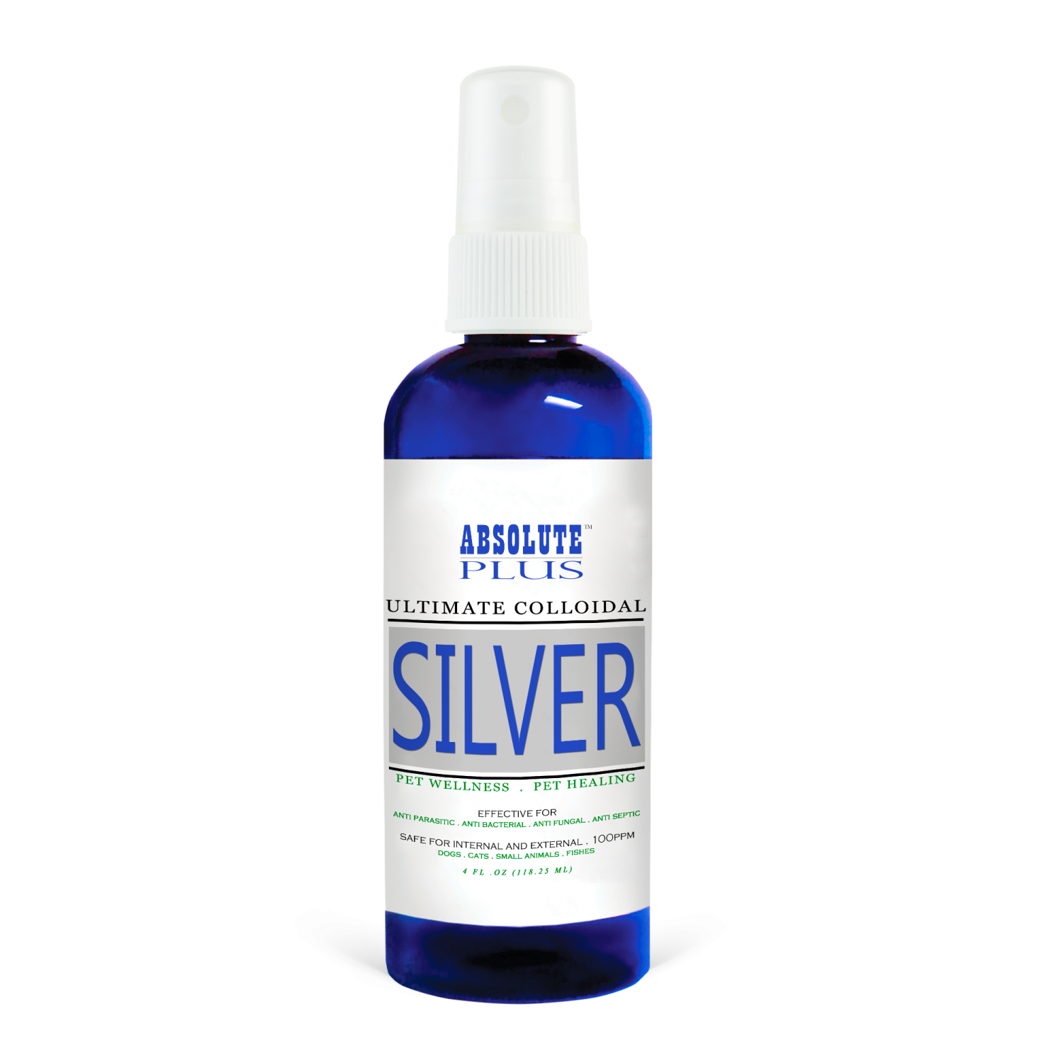
Ultimate Colloidal Silver » Absolute Pets
Colloidal Silver 100ml (Higher Nature) - HealthySupplies.co.uk. Buy Online.
Dom & Cleo Colloidal Silver For Dogs & Cats (2 Sizes) | PerroMart Singapore – PerroMart SG
Five Immune-Boosting Uses of Colloidal Silver | Dogs Naturally
Colloidal Silver Liquid - 40 PPM Colloidal Silver Liquid Manufacturer from Vadodara
Colloidal Silver... Nature's Healer - Marsillo Chiropractic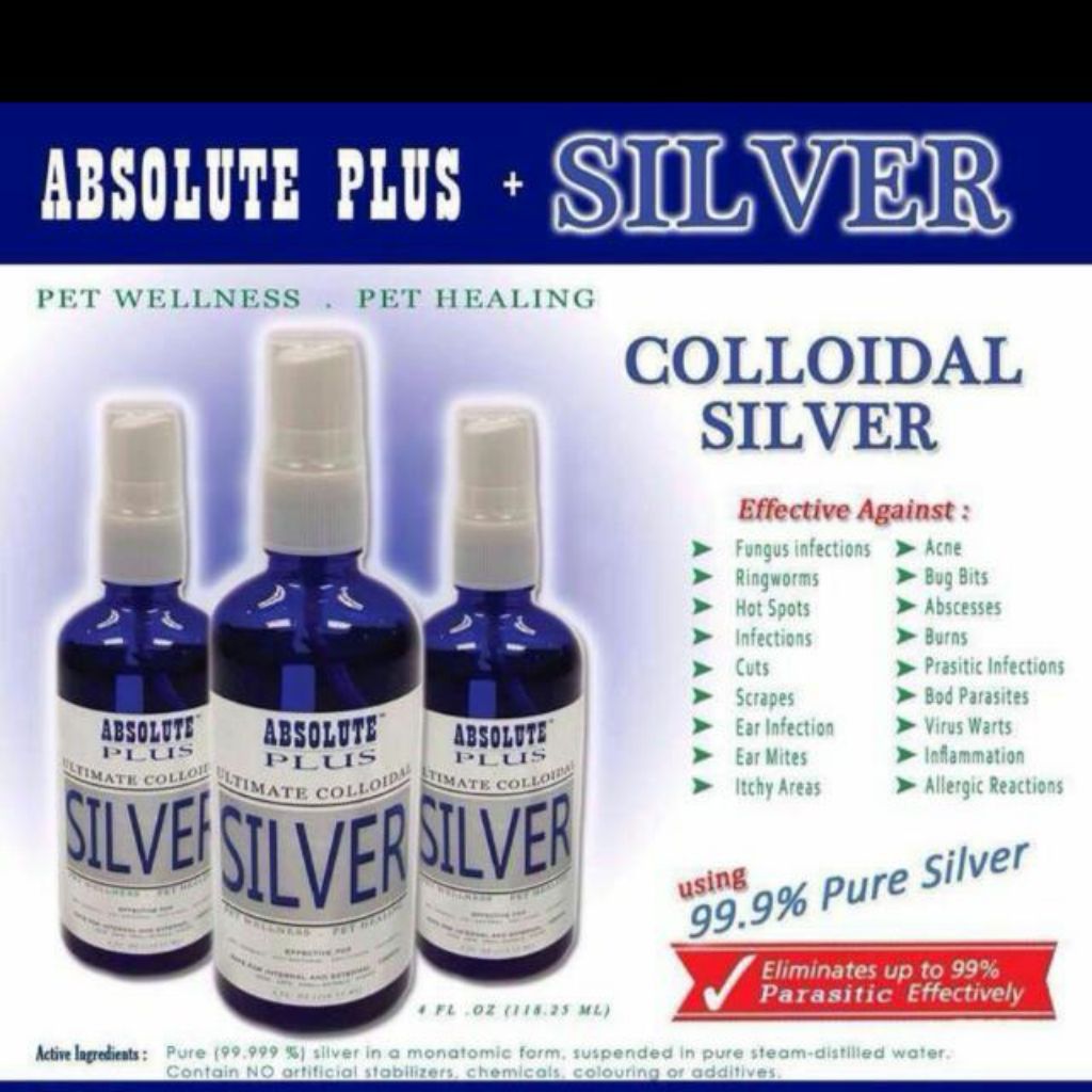
Shopee Singapore | Buy Everything On Shopee
GU GUIDE Bali - WELLNESS WEDNESDAY: COLLOIDAL SILVER Silver, in its many forms that include chelated, nano, ionic, and colloidal silver, is gaining ground as more people realize the very real antibiotic
The silver lining: towards the responsible and limited usage of silver - Naik - 2017 - Journal of Applied Microbiology - Wiley Online Library
Toxicity of colloidal silver products and their marketing claims in Finland - ScienceDirect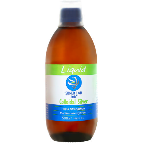
Silverlab Ionic Colloidal Silver Liquid 500ml - Clicks
Colloidal Silver: Uses, Benefits and Dangers.jpg)
The Applications of Colloidal Silver
Colloidal Silver For Dogs: A Natural Antibiotic for Sick Dogs | CertaPet
Silver Lab Colloidal Silver 100ml | Online Shopping | Wellness Warehouse
Colloidal Silver | Book by Werner Kühni, Walter von Holst | Official Publisher Page | Simon & Schuster


.jpg)


Posting Komentar untuk "colloidal silver for yeast infection"